Endometriosis surgery: comparing excision and ablation paths
The choice between ablation and excision shapes what surgery can accomplish, but the difference is not simply that one approach treats the surface and the other reaches the whole lesion.
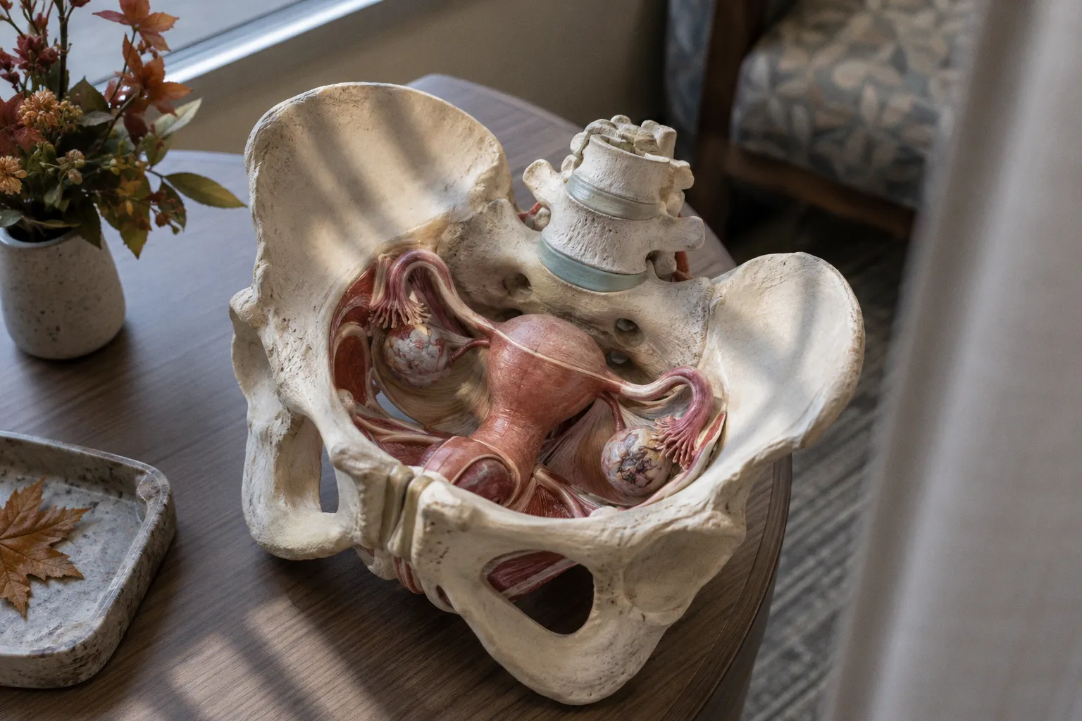
Ablation uses energy to destroy targeted tissue; how deeply that effect extends depends on the technique, equipment, tissue, and surgical judgment. Excision removes tissue for examination and aims to take out the lesion, though complete removal can be difficult when disease is extensive or close to critical structures.
For ovarian endometriomas, a 2024 Cochrane review reported recurrence at one year in 37% of patients after ablation and 5–17% after excision. Those figures make the comparison worth taking seriously. They do not, on their own, predict an individual patient’s pain, fertility, or recovery. The location and extent of disease, the procedure performed, and the experience of the surgical team all matter.
How the two operations work
Ablation applies energy to an endometriosis lesion to destroy tissue. Surgeons may use electrosurgery, laser, plasma energy, or other energy-based methods. Treatment is directed at the visible lesion, but its depth is variable. It can extend below the surface, with the effect shaped by the modality and how it is applied.
Excision removes a portion of tissue with instruments such as scissors or a cold loop. The surgeon separates the lesion from surrounding structures and removes it, sometimes with a margin of nearby tissue. The amount that can be safely removed depends on the lesion’s borders, the surrounding anatomy, and whether disease involves organs such as the bowel, bladder, or ureter. Ultrasonic instruments, including harmonic shears, use energy and should not be described as cold-cutting tools.
The distinction matters, but neither technique guarantees that every diseased cell has been treated. Endometriosis can extend beneath the visible surface, and scar tissue can obscure its borders. Ablation can destroy tissue below the area seen at laparoscopy; excision can provide a specimen, but the completeness of removal depends on what the surgeon can safely identify and remove.
Ablation destroys targeted tissue in place. Excision removes tissue for examination, when it can be safely removed.
The procedures also differ in what happens after the operation. Ablation generally leaves no intact specimen from the treated area. Excision usually does, allowing a pathologist to examine the removed tissue. That distinction can inform diagnosis, but it should not be confused with a guarantee that excision is appropriate for every lesion or every patient.
Recurrence, pain, and long-term outcomes
The 2024 Cochrane review found a difference in one-year ovarian endometrioma recurrence: 37% after ablation and 5–17% after excision. These results concern endometriomas, not every form of endometriosis. They also describe recurrence in a group of patients, not the outcome any one person should expect.
| Consideration | Ablation | Excision |
|---|---|---|
| What the surgeon does | Applies energy to destroy targeted tissue | Removes tissue with instruments such as scissors or a cold loop |
| Depth of treatment | Variable; may extend beneath the visible surface | Depends on the tissue removed and what can be safely separated |
| Tissue for pathology | Usually no intact specimen from the treated area | Removed tissue can generally be sent for histology |
| One-year ovarian endometrioma recurrence in the 2024 Cochrane review | 37% | 5–17% |
| Suitability for complex disease | Depends on site, depth, anatomy, and expertise | May require advanced skills and a multidisciplinary team |
Pain returning after surgery does not establish why it has returned. Possible explanations include persistent disease, recurrence, adhesions, pelvic floor dysfunction, or another condition that can cause pelvic pain. Nor does pain returning soon after surgery prove that ablation left disease behind. A careful assessment looks at the original findings, the operation performed, symptom pattern, examination, and, when appropriate, imaging.
Long-term pain management may involve more than another operation. Hormonal treatment, pelvic floor physical therapy, pain-focused care, and support for fertility goals can all be part of a plan. The right combination depends on symptoms and priorities. Surgery is one part of care, not a promise that pain will disappear permanently.
Histology: what a specimen can and cannot confirm
When tissue is removed, it can be examined under a microscope. Histology may confirm endometriosis by identifying characteristic glands and stroma. It can also help distinguish endometriosis from other conditions. Ablation usually does not provide an intact specimen from the treated lesion, which limits what can be confirmed about that specific tissue.
Histology is useful, but it is not the only way clinicians assess endometriosis. Imaging, symptoms, examination, and findings during surgery all contribute to diagnosis and treatment planning. A negative or unavailable pathology result does not by itself settle whether a patient has endometriosis, just as a positive specimen does not explain every source of pain.
For a patient who has had years of pelvic pain without a clear diagnosis, a pathology result may provide valuable information. It can guide follow-up and future discussions with clinicians. Its role should be described plainly: it documents what was found in the tissue sampled, while the wider clinical picture still matters.
Deep infiltrating endometriosis and surgical complexity
Deep infiltrating endometriosis can involve the bowel, bladder, ureters, or structures along the pelvic sidewall. In these locations, the central issue is not just whether to use energy or remove tissue. It is whether the disease can be treated safely, what organs are involved, and whether the surgical team has the expertise and support the case requires.
Energy applied near bowel, ureters, nerves, or blood vessels carries risks that vary with the location and depth of disease. Excision in these areas can also be complex. Depending on the findings, surgery may involve freeing a ureter from surrounding tissue, removing a portion of the bladder, or working with a colorectal surgeon if bowel disease is present. Not every patient with suspected deep disease needs these procedures, and not every operation can remove all visible disease without unacceptable risk.
A thorough preoperative plan matters. Imaging may help map disease and identify the teams needed, though it cannot answer every question about what will be found during surgery. Patients can ask whether the surgeon regularly treats deep disease, what procedures might be needed if it involves an organ, and which specialists would be available. Clear answers help set realistic expectations about both the operation and recovery.
The choice of surgeon matters particularly when endometriosis is deep or extensive. A laparoscopic procedure can be minimally invasive in its access, yet technically demanding in what it asks the surgeon to do. If the scope of disease is uncertain, referral for specialist assessment can help clarify options before consent.
Recovery and ongoing care
Recovery after minimally invasive gynecologic surgery depends on what was done, not just the name of the procedure. A limited operation on superficial disease may have a different course from ovarian cyst surgery or dissection around bowel or ureters. Patients should ask their own surgical team for guidance on lifting, driving, work, exercise, and when to seek help. A single timetable cannot account for these differences.
Some fatigue, bloating, and discomfort can occur during recovery. Symptoms should gradually be considered in the context of the operation and the patient’s overall progress. Worsening pain, new or persistent bowel or bladder problems, fever, or other concerning changes warrant contact with the care team rather than relying on a generic recovery schedule.
Long-term care is often part of the surgical plan. Hormonal suppression, including progestins, combined hormonal contraception, or GnRH-based treatment, may be discussed to help manage symptoms and reduce the risk of recurrence. The choice depends on medical history, side effects, and whether pregnancy is a current goal. Suppression does not guarantee that disease will not return, and it is not compatible with trying to conceive while in use.
Follow-up should make room for changing priorities. A patient’s plan may need to shift as pain, medication tolerance, or fertility goals change. Imaging may be appropriate for some patients, particularly when ovarian or deep disease was part of the original picture, but the timing and need should be individualized. People trying to conceive may need a fertility discussion sooner than those whose immediate priority is symptom control.
The most useful question is not which operation is universally best. It is which approach fits the location and extent of disease, the goals of treatment, and the expertise available. Excision offers tissue for pathology and, for ovarian endometriomas, lower one-year recurrence in the Cochrane review cited here. Ablation can be appropriate in selected circumstances, but its effects are not confined automatically to the visible surface. A sound plan explains what the surgeon expects to treat, what may remain uncertain, and how care will continue after the incisions heal.